Can Craniopharyngioma Be Linked To Military Service
Case report
peer-reviewed
Hypothalamic Injury Post-obit Surgery for Craniopharyngioma Causing Immediate Postoperative Decease
Abstract
While autonomic disturbances resulting from a hypothalamic injury are uncommon complications following surgery for craniopharyngioma, they can lead to postoperative death. Herein, we discuss the case of a multicompartmental craniopharyngioma in a 13-year-old kid who died due to unexpected hypothalamic injury, resulting in rapid deterioration in the hemodynamic and neurological status of the patient.
Introduction
Craniopharyngiomas are rare, beneficial sellar/parasellar tumors derived from embryonic tissue [1]. Even though these tumors are slow-growing and beneficial, they cause complications every bit a issue of the mass outcome on the neighboring structures. This results in the patient presenting with symptoms, such as headaches, bitemporal hemianopia, and hypopituitarism.
Radical excision of craniopharyngiomas normally leads to injury to the hypothalamus. Nonetheless, complications arising from autonomic dysfunction, which was seen in our case, are uncommon and do not respond well to treatment.
Case Presentation
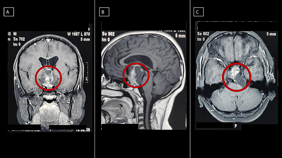
A 13-twelvemonth-old child was admitted with an eight-month history of headache and recurrent seizures. Neurological exam was unremarkable with no motor or sensory deficits preoperatively. There was, all the same, bilateral papilledema. MRI was done which revealed that the tumor was multicompartmental, extending from the suprasellar cistern to the right subtemporal region and invading the prepontine cistern, loosely adherent to the basilar avenue (Figure i).
Under the cover of antibiotics, anti-epileptics, and mannitol, a left frontotemporal craniotomy, also known as pterional craniotomy, was done. A transsylvian approach was taken, the arachnoid affair was opened, and the right internal carotid artery and optic nerve were identified. Then, via the optico-carotid cistern, a gross full resection (GTR) of the tumor was performed with sufficiently condom margins to avoid the take a chance of recurrence. Except for the hypothalamus, at that place was a good cleavage plane from the surrounding neural and vascular structures and the tumor.
The whole operative procedure was uneventful. The patient was hemodynamically stable with a normal sensorium immediately after surgery. The only abnormalities noticed were right lateral rectus weakness concomitant with subtle right-sided pyramidal weakness.
On the fifth 24-hour interval, he developed diabetes insipidus with a serum sodium concentration of 165 mEq/L and an increased urine output of approximately ii.5 L - 3 50 per day. He was treated with desmopressin spray and intravenous fluids. On the evening of postoperative Day vii, the patient started developing hypothermia with a temperature of 33°C and started shivering. Arterial blood gas (ABG) analysis was done frequently and was establish to be normal. A postoperative CT scan of the brain showed a left frontotemporal craniotomy defect with postoperative changes at the sellar and suprasellar region and minimal residual tumor with perilesional edema.
The next morning, he was found to exist in a Glasgow Coma Scale (GCS) of 7 which required immediate intubation and ventilation. The next day, the patient developed wide claret pressure fluctuations. An echocardiogram identified a left ventricular dysfunction. Soon after, despite the resuscitative measures, the patient succumbed to a fatal cardiac arrest.
Discussion
Despite all of the contempo advances in operative techniques and surgical equipment, craniopharyngioma is considered difficult to treat; the optimal handling strategy for it remains ambiguous [2].
A craniopharyngioma is a benign tumor, GTR is considered to exist the gold standard. However, this approach can be challenging due to the location of these tumors, which is usually in close proximation with structures, such as the optic chiasm, hypothalamus, and internal carotid arteries [3-4]. A more bourgeois approach is a subtotal resection (SR) with subsequent radiation therapy (RT) which consists of deliberately leaving around 10% of the residual lesion. This approach has been shown to take reduced surgical complications, but there are higher chances of tumor recurrence and agin effects from radiations [5-half-dozen].
Postoperative morbidities due to GTR unremarkably encompass endocrinopathies leading to panhypopituitarism; withal, death as a result of hypothalamic damage is rarely reported [ii, 7]. Clinical manifestations due to the destruction of various regions of the hypothalamus are illustrated in Effigy 2.
Based on the clinical characteristics of the patient, it can be hypothesized that the following areas of the hypothalamus were damaged: supraoptic and paraventricular nuclei (causing diabetes insipidus) as well every bit the posterior hypothalamus, ventromedial, lateral, paraventricular, and anterior nuclei (causing hypothermia, tachycardia, and wide fluctuations in blood pressure). The cardiac dysfunction was a result of the injury to the autonomic region of the hypothalamus resulting in ventricular failure and somewhen death.
It is our belief that our attempt at GTR proved to be fatal due to the damage to multiple hypothalamic nuclei. This injury to the hypothalamus may have been a result of direct trauma to it or an infarction following injury to the vessels that supply the hypothalamus. A less radical approach, such equally SR with RT, would have probably avoided the patient's mortality, especially when we noticed multiple hypothalamic nuclei adherent to the surface of the tumor. This is farther substantiated by a systematic review done by Yang et al. that showed that SR + RT was an acceptable approach to accomplish tumor control while limiting hypothalamic morbidity associated with GTR [8].
Conclusions
GTR is considered to be the gold standard handling for craniopharyngioma and is successful in most cases equally information technology hinders the chances of recurrences of the tumor. Yet, in our instance, the tumor was noted to be multi-compartmental and was adherent to multiple nuclei of the hypothalamus. The use of GTR in such circumstances led to damage of various nuclei of the hypothalamus, resulting in deterioration in the patient'due south health and, finally, led to his demise. We conclude that, in cases such equally ours, the idea of SR followed by RT may show to be a amend approach despite having a higher recurrence rate considering the life-threatening farthermost damage to the hypothalamic nuclei can be avoided.
References
- Karavitaki Due north, Cudlip S, Adams CB, Wass JA: Craniopharyngiomas. Endocr Rev. 2006, 27:371-97. 10.1210/er.2006-0002
- Weiner HL, Wisoff JH, Rosenberg ME, et al.: Craniopharyngiomas: a clinicopathological analysis of factors predictive of recurrence and functional effect. Neurosurgery. 1994, 35:1001-ten. 10.1227/00006123-199412000-00001
- Hofmann BM, Höllig A, Strauss C, Buslei R, Buchfelder One thousand, Fahlbusch R: Results after treatment of craniopharyngiomas: further experiences with 73 patients since 1997. J Neurosurg. 2012, 116:373-84. 10.3171/2011.vi.JNS081451
- Morisako H, Goto T, Goto H, Bohoun CA, Tamrakar S, Ohata K: Aggressive surgery based on an anatomical subclassification of craniopharyngiomas. Neurosurg Focus. 2016, 41:E10. x.3171/2016.9.FOCUS16211
- Clark AJ, Muzzle TA, Aranda D, Parsa AT, Sun PP, Auguste KI, Gupta Due north: A systematic review of the results of surgery and radiotherapy on tumor control for pediatric craniopharyngioma. Childs Nerv Syst. 2013, 29:231-38. 10.1007/s00381-012-1926-2
- Burman P, van Beek AP, Biller BM, Camacho-Hübner C, Mattsson AF: Radiotherapy, especially at immature age, increases the gamble for de novo brain tumors in patients treated for pituitary/sellar lesions. J Clin Endocrinol Metab. 2017, 102:1051-58. 10.1210/jc.2016-3402
- Pereira AM, Schmid EM, Schutte PJ, et al.: High prevalence of long-term cardiovascular, neurological and psychosocial morbidity afterwards treatment for craniopharyngioma. Clin Endocrinol (Oxf). 2005, 62:197-204. x.1111/j.1365-2265.2004.02196.10
- Yang I, Sughrue ME, Rutkowski MJ, et al.: Craniopharyngioma: a comparison of tumor control with various handling strategies. Neurosurg Focus. 2010, 28:E5. 10.3171/2010.1.FOCUS09307
Instance report
peer-reviewed
Hypothalamic Injury Post-obit Surgery for Craniopharyngioma Causing Immediate Postoperative Decease
Ethics Statement and Conflict of Interest Disclosures
Human subjects: Consent was obtained or waived past all participants in this study. Conflicts of interest: In compliance with the ICMJE uniform disclosure form, all authors declare the following: Payment/services info: All authors have declared that no financial support was received from any organisation for the submitted piece of work. Financial relationships: All authors have alleged that they have no financial relationships at present or within the previous three years with whatsoever organizations that might have an interest in the submitted piece of work. Other relationships: All authors have declared that there are no other relationships or activities that could appear to have influenced the submitted piece of work.
Article Information
DOI
10.7759/cureus.18814
Cite this article every bit:
Bhandari P, Nagpal S, Parthasarathi A, et al. (October 16, 2021) Hypothalamic Injury Following Surgery for Craniopharyngioma Causing Immediate Postoperative Decease. Cureus 13(10): e18814. doi:10.7759/cureus.18814
Publication history
Peer review began: September 26, 2021
Peer review concluded: October 12, 2021
Published: October 16, 2021
Copyright
© Copyright 2021
Bhandari et al. This is an open up admission article distributed nether the terms of the Artistic Eatables Attribution License CC-By 4.0., which permits unrestricted utilise, distribution, and reproduction in whatsoever medium, provided the original author and source are credited.
License
This is an open up admission commodity distributed under the terms of the Creative Commons Attribution License, which permits unrestricted use, distribution, and reproduction in any medium, provided the original writer and source are credited.
Instance report
peer-reviewed
Hypothalamic Injury Following Surgery for Craniopharyngioma Causing Firsthand Postoperative Death
Figures etc.
Can Craniopharyngioma Be Linked To Military Service,
Source: https://www.cureus.com/articles/72963-hypothalamic-injury-following-surgery-for-craniopharyngioma-causing-immediate-postoperative-death
Posted by: mcpeekhurse1984.blogspot.com


0 Response to "Can Craniopharyngioma Be Linked To Military Service"
Post a Comment